Research highlights 💡
My research fits in the broader theme of multiscale connectomics of the healthy and epileptic brains. Below, you will find a subset of research projects, reviews, and toolbox I have published. Hover over the figures to find out more!
The ENIGMA Toolbox
An open-source ecosystem for accessing 100+ ENIGMA statistical maps, visualizing cortical and subcortical surface data, relating neuroimaging findings to micro- and macroscale brain organization, and so much more! Check out our documentation!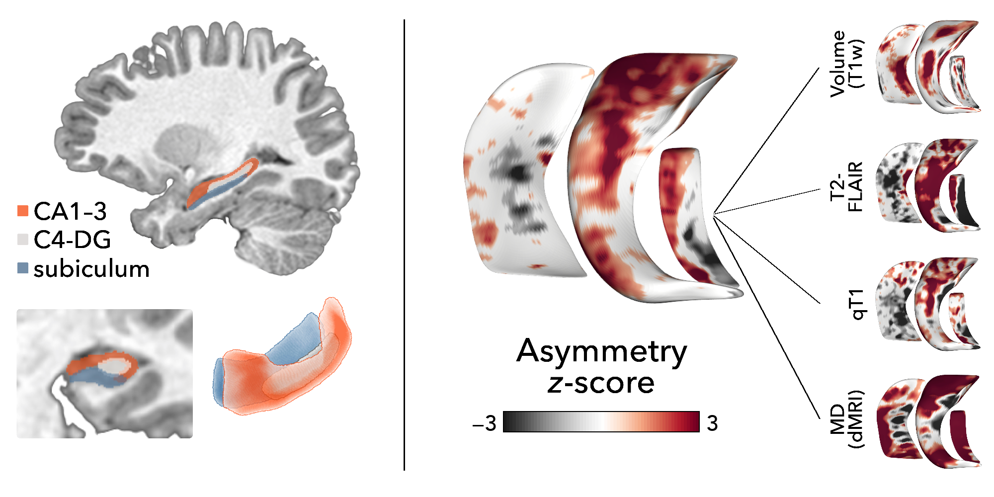
Case study in drug-resistant childhood epilepsy
Together with the ILAE Neuroimaging Task Force, this educational case report advocates for the clinical relevance of quantitative MRI markers of hippocampal pathology for diagnosis and surgical target identification in pediatric drug-resistant epilepsy.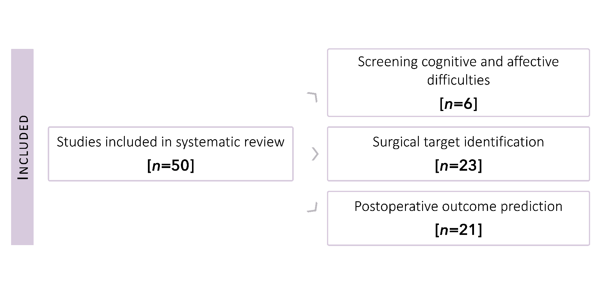
Biomarkers of drug-resistant epilepsy
This systematic review highlights previous neuroimaging biomarkers of drug-resistant epilepsy, focussing on three key application areas: (i) prediction of cognitive impairments, (ii) lesion detection, and (iii) postoperative outcome prognosis.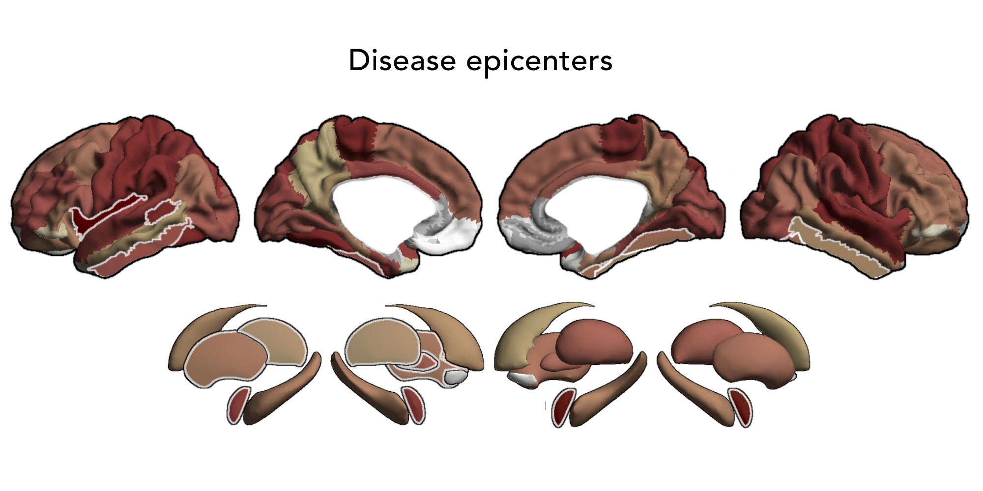
Network models in epilepsy
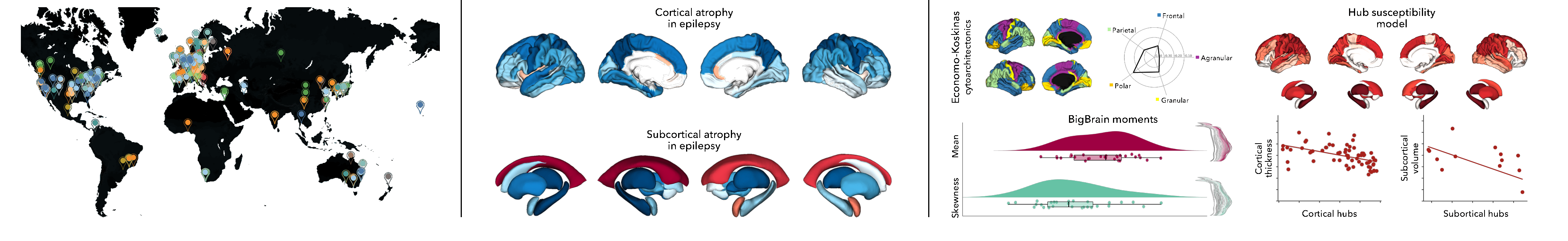
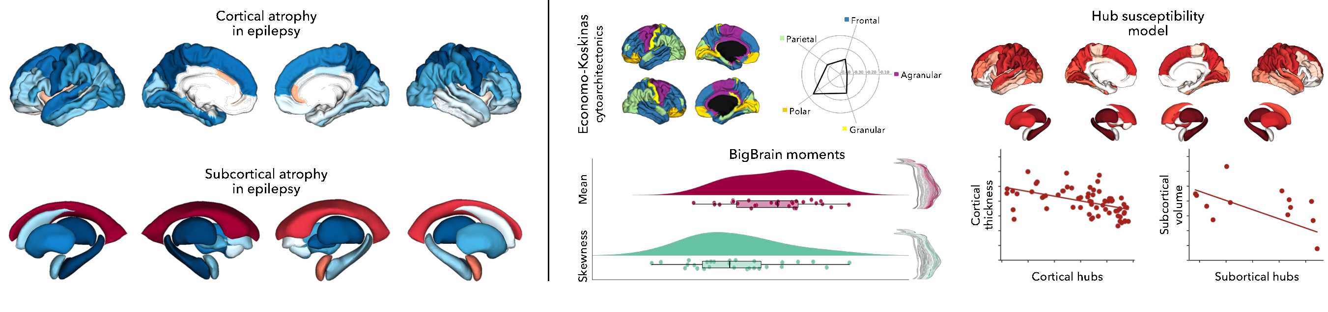
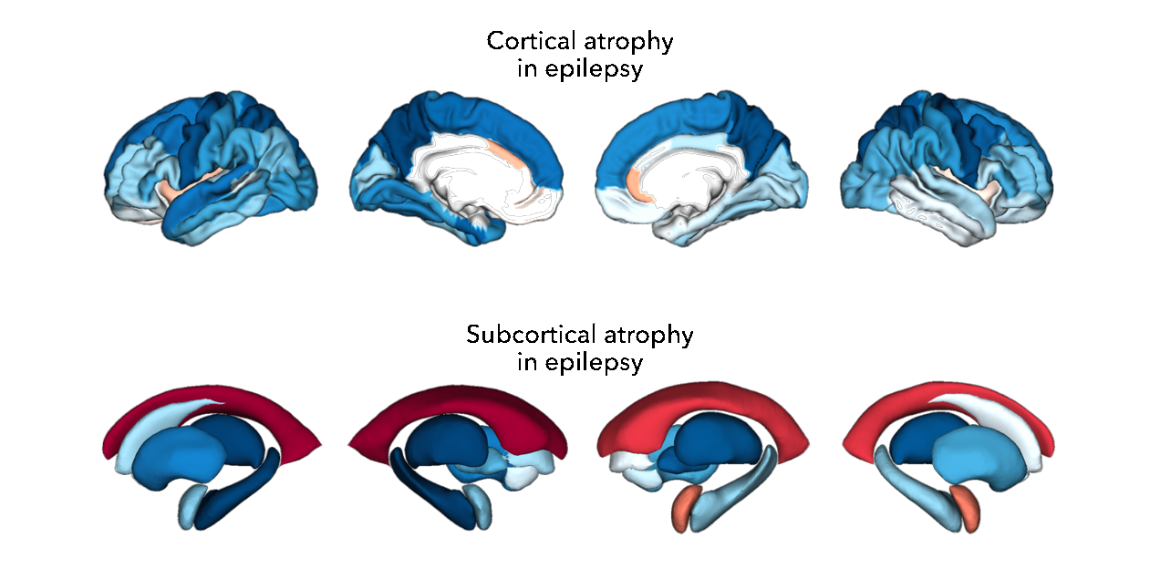
Through worlwide collaboration in ENIGMA-Epilepsy, we showed that epilepsy-related atrophy patterns co-localized with hub regions and were anchored to the connectivity profiles of distinct subnetworks. Together, these findings provided deeper insights into the macroscale features that shape the pathophysiology of common epilepsies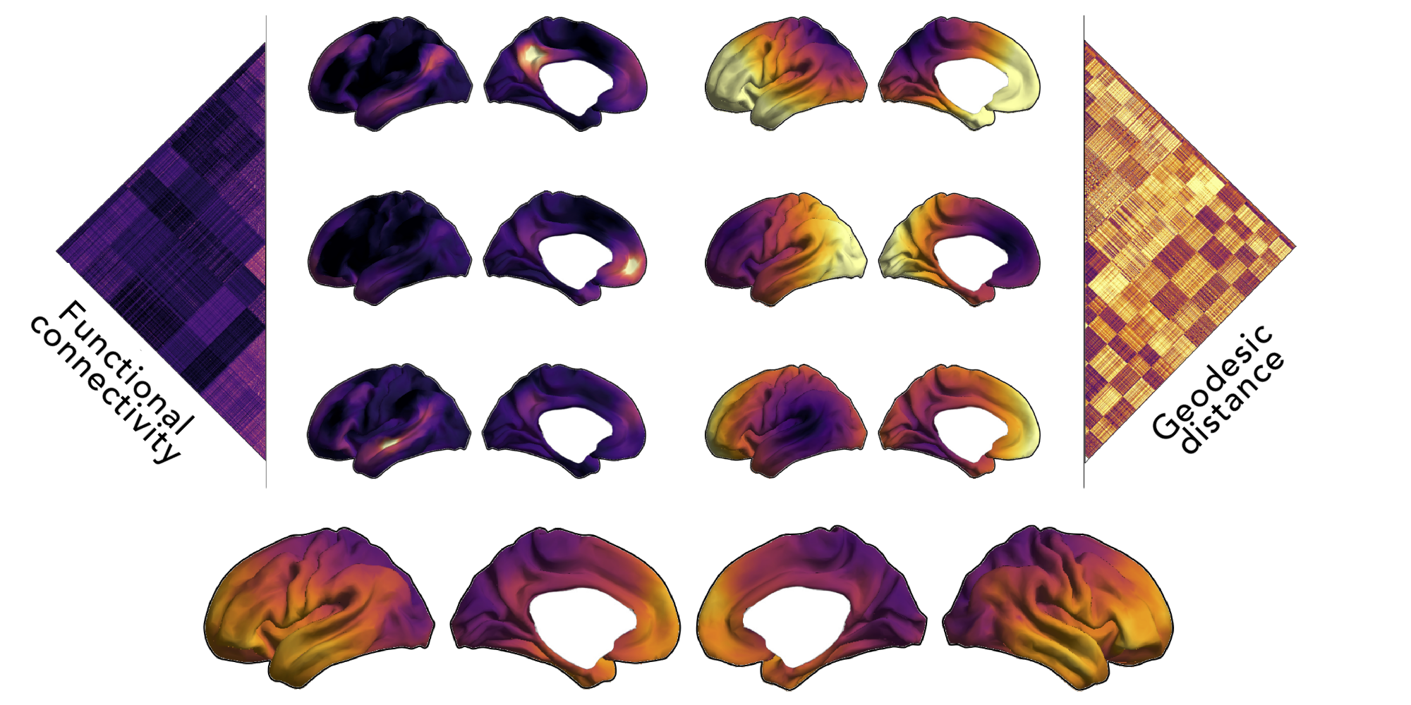
Connectome contractions in epilepsy
By combining functional connectivity and geodesic distance, we showed that patients with temporal lobe epilepsy had imbalances in short- vs. long-range connections, which were independent of atrophy but mediated by white matter changes. A machine learning algorithm could predict postsurgical outcome from connectivity distance features with 76% accuracy.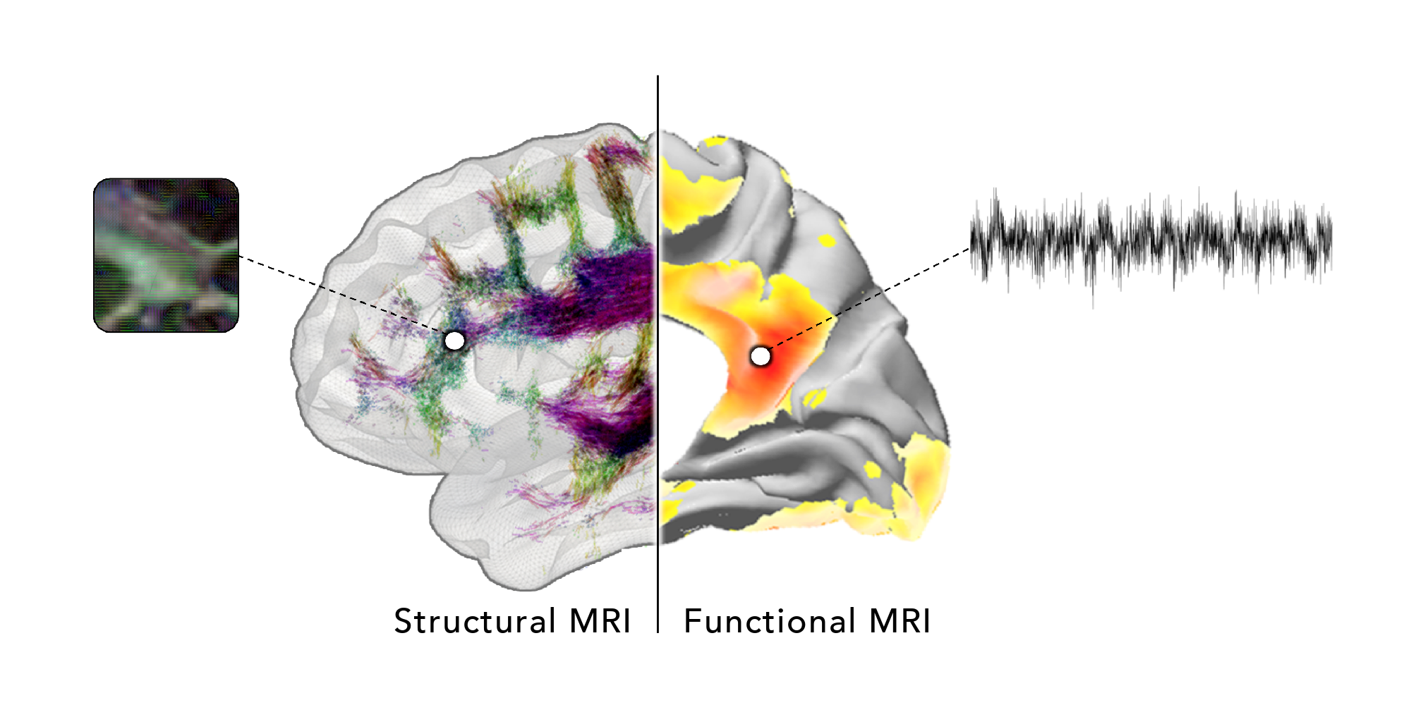
Microstructure-informed connectomics
In this review, we argue for a science of the brain that embraces features along a continuum between microscale (gene expression), mesoscale (morphology), and macroscale topology (connectomes). We also discuss how this approach can improve our understanding of prevalent brain disorders.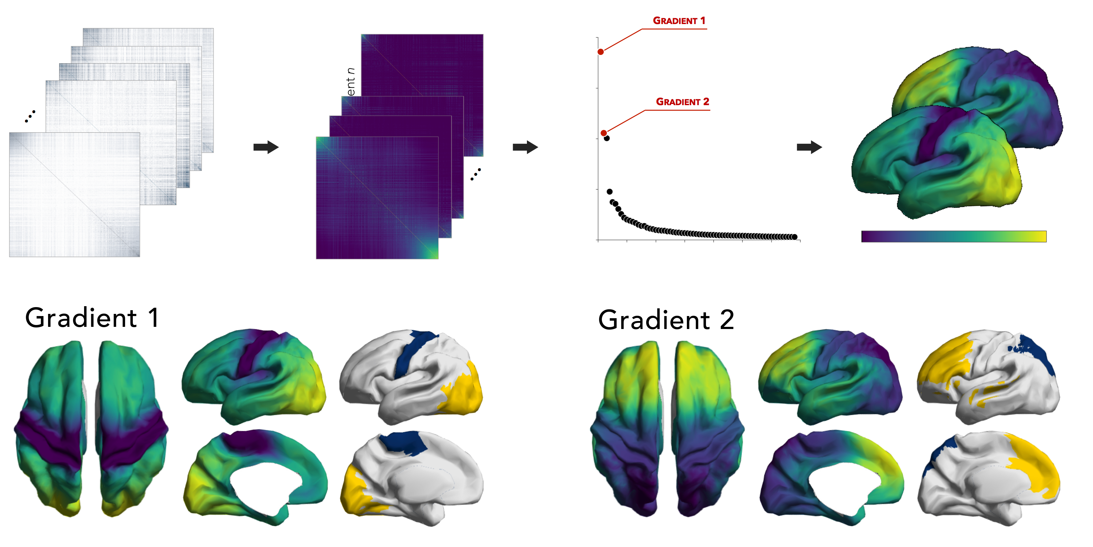
Baby gradients
Our investigation into functional gradients—and their correspondence to multiscale features—in neonates revealed a gradient running from sensorimotor to visual cortices, capturing associations to white matter microstructure and thalamo-cortical connectivity, as well as a gradient depicting an immature differentiation between unimodal and transmodal areas.I am grateful for funding from the Fonds de recherche – Santé (FRQS) and the Canadian Institutes of Health Research (CIHR).
![]()
![]()
© SL 2023